Relevant Skills
Image Processing (MATlab)
Biological Sample Handling
Protocol Design
SLA Printing
Design for Research Environment
Motivation
Biological research benefits from the existence of high complexity in vitro models – the better the in vitro model is, the greater the scientists ability to experiment in more accurate conditions prior to involving live samples. Organoids are one such model, these three dimensional, multicellular, self organizing masses are the basis for of a rapidly expanding field. In essence, stem cells are guided through the differentiation process to produce complex tissues such as the following:
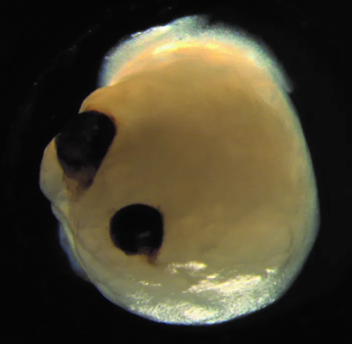
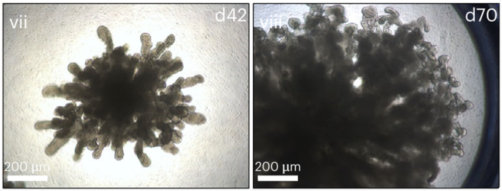
As is clear in these images, these tissues introduce a new imaging problem, we cannot see inside them. There are many ways to work around this limitation, some labs opt for sacrificing their samples, others choose to focus on exterior traits, and some have looked to optical coherence tomography (OCT) to provide 3D imaging. However, some of these tissues are inherently load bearing or structural tissues such as cartilage, and require mechanical analysis, which OCT cannot provide. This, again, results in the need to sacrifice the sample, slice into thin segments, then subject them to traditional shear or torsional testing machinery.
Our proposition is to utilize Optical Coherence Elastography to measure the shear modulus of 3D tissues nondestructively.
Optical Coherence Elastography (OCE)
OCE uses OCT to visualize the motion of shear waves across a material. In essence, a high frequency shear wave is excited in the tissue of interest, then we scan twice over each location and compare the measured waves (light) to each other, and use the shift to calculate the distance that portion of tissue has moved. This can then be used to calculate the speed of the tissues vibration. For a more math forward explanation feel free to look at this paper, which my lab published recently and is the basis of the processing pipeline we currently use.
System Design & Results
The core constraint for this system is the ability to excite high enough frequency shear waves to observe one and a half or so periods within the tissue. This difficulty arises from the nature of shear wave propagation:
As tissue stiffness \( \mu_s\) increases, for the same density (which we generally estimate to equal the density of water) we see an increase in shear wave speed propogating through the material.
As shear wave speed increases as a result of stiffer tissues, and wave length must decrease as a result of smaller tissues, we are required to push frequency higher.
For similar applications in the eye we need a minimum of 1-2kHz of excitation, for the cartilage organoids we will be working with , we need a minimum of 6-7 kHz of excitation. These high frequency waves will attenuate much more quickly than their low frequency counterparts, introducing the need for higher amplitude excitation as well.
Design Requirements
- Compatible with various sample shapes and sizes.
- Compatible with convenient imaging mediums for high throughput (non disruptive).
- Piezoelectric device must be protected from small amounts of liquid.
Unfortunately this is all I can share until we complete our patent submission! In the meantime this abstract outlines the system results: